
Explore our diverse research areas, groundbreaking studies and remarkable medical research achievements
A study published in Cell on July 20, 2026, presents Viral protease-Initiated lytic cell Death (VID), a universal antiviral mRNA therapeutic platform that exploits viral proteases to selectively destroy infected cells. The work was carried out jointly by Aiping Wu’s group at the Suzhou Institute of Systems Medicine (CAMS & PUMC) and Cheng-Feng Qin and Yi-Jiao Huang’s group at the Academy of Military Medical Sciences (AMMS). The study introduces the concept of VID and demonstrates that a single modular platform, combined with a generative AI–driven molecular design workflow, can target phylogenetically diverse viruses, completing the full design-to-validation cycle in one round of experiments.
Viruses depend on their own proteases to cleave polyproteins early in infection, and these enzymes are highly selective for their cognate substrates. VID activators (VIDAs) turn this selectivity against the virus. The native caspase-cleavage site in the pyroptotic effector GSDMD is replaced with a viral protease recognition motif. As a result, the protease that the virus must produce to replicate now also activates GSDMD, triggering pyroptosis in infected cells while leaving uninfected bystanders untouched. The choice of pyroptosis, rather than apoptosis, is deliberate: the lytic, pro-inflammatory nature of pyroptotic cell death releases damage signals that help recruit and activate downstream immune responses. To prevent inadvertent activation by host caspases, six additional residues in GSDMD were mutated to abolish endogenous recognition - a crucial safeguard for uninfected bystander cells.
The team first constructed a VIDA responsive to the HAV 3C protease. In cell culture, activation was strictly protease-dependent: uninfected cells were unharmed, and infected cells were efficiently lysed. In HAV-infected mice, intravenous delivery of LNP-encapsulated VIDA mRNA reduced fecal viral loads by approximately two orders of magnitude (~100-fold) and drove hepatic viral RNA to near the limit of detection. In contrast, control animals showed the hallmarks of liver inflammation — elevated transaminases and immune-cell infiltration — that were absent or markedly reduced in VIDA-treated mice. The treatment remained effective even when initiated four days post-infection.
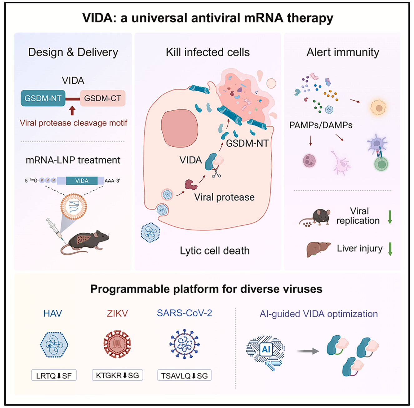
The mechanism of VIDA
Single-cell transcriptomics revealed that the pyroptotic response went beyond eliminating infected hepatocytes. Dying cells released inflammatory signals that drew immune cells into the liver: interferon and antigen-presentation pathways were upregulated, and both CD8⁺ effector and CD4⁺ helper T cells expanded. This “kill-and-alert” mechanism means VIDA acts simultaneously as a therapeutic and an immunogen, clearing the virus while priming adaptive immunity. In both single- and repeat-dose regimens, the construct showed no signs of liver or kidney toxicity and no cytokine storm.
VIDA is modular by design: swapping the protease-recognition cassette redirects the platform to a different pathogen. The team demonstrated this for the Zika virus (ZIKV) NS2B/3 protease and the SARS-CoV-2 main protease NSP5. Both were validated in cell-based assays; the ZIKV construct was further confirmed in an animal model. That the same scaffold works against three virus families as phylogenetically diverse as Picornaviridae, Flaviviridae, and Coronaviridae illustrates the breadth of the approach, limited mainly by the requirement for a virus-encoded protease with defined substrate specificity.
Optimizing the SARS-CoV-2 cleavage motif ran into a practical bottleneck: natural cleavage-site sequences were plentiful, but their kinetic activities remain largely uncharacterized, and experimental screening was expensive. Rather than iterate through rounds of trial and error, the team devised a generate–sample–validate workflow built on an evolutionary model of variant effect (EVE, a variational autoencoder). Trained on just 261 non-redundant natural NSP5 cleavage motifs, the model learned to distinguish functional from non-functional sequence space. On an independent test set, its evolutionary scores discriminated cleavable from non-cleavable motifs with an ROC-AUC of 0.80.
From this learned space, 100,000 candidate sequences were sampled and passed through clustering-based diversity selection, off-target screening, and toxicity filtering, leaving 30 candidates for synthesis, of which 25 were successfully produced. Cleavage rates ranged widely, from near-zero to approximately 16-fold above wild type, with seven candidates showing >2-fold higher cleavage rates than wild type. Evolutionary scores correlated significantly with measured cleavage activity (Spearman, p < 0.001).
When installed in the VIDA backbone, six of the seven AI-designed molecules drove stronger lysis than the parental construct. The three best (variants #1, #3, and #4) suppressed SARS-CoV-2 replication by over 90% in infected cells. Kinetic analysis showed that the improvement came from faster catalytic turnover (k cat) rather than tighter substrate binding (K m): the AI had found sequences that are cleaved faster, not ones that grip the protease more tightly. From model training to functional validation, the entire cycle required only a single round of wet-lab experiments.
The central insight of VIDA is that a viral protease — the one enzyme a virus cannot afford to lose — can be turned into the trigger for its host cell’s death. Because the platform is modular and delivered as mRNA, it pairs rapid manufacturing with rapid retargeting: in principle, any virus that encodes a protease with defined substrate specificity is a candidate. The generative AI workflow further compresses the timeline by replacing iterative screening with computationally guided, single-round optimization. Together, mRNA production speed and AI-driven design offer a path toward broad-spectrum antivirals that can be redirected to a novel pathogen within the time it takes to characterize its protease, without starting drug development from scratch.
Cheng-Feng Qin, Yi-Jiao Huang, and Aiping Wu are co-corresponding authors. Lin Li, Xiu-Li Yan, Hao-Yang Wang, Hang-Yu Zhou, and Yu-Yan Li are co-first authors. The study was supported by the National Key Research and Development Program of China, the National Natural Science Foundation of China, the CAMS Innovation Fund for Medical Sciences, and other funding sources.
Reference: Li et al., Viral protease-initiated lytic cell death as a universal antiviral mRNA therapy. Cell189(17), 5396–5410.e8 (2026). https://doi.org/10.1016/j.cell.2026.06.031

