
Explore our diverse research areas, groundbreaking studies and remarkable medical research achievements
Source: Chinese Academy of Medical Sciences & Peking Union Medical College — Institutional News (report from Fuwai Hospital)
Original URL: https://www.pumc.edu.cn/pub/cams/yxbd/422b6552aeb54ef0ac5ee1e96c5336ef.htm
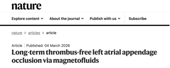
Fuwai Hospital of the Chinese Academy of Medical Sciences, in collaboration with the Chinese Academy of Sciences, has released a magnetofluid-based intracardiac 3D printing technology, published in the top international journal Nature.
Introduction: A Paradigm Revolution from "Metal Devices" to "Fluid Shaping"
Background
As population ageing intensifies, atrial fibrillation has become a highly prevalent cardiovascular disease worldwide, with a global patient total of 60 million. The most lethal complication of atrial fibrillation is stroke (cerebral embolism). Clinical research has confirmed that about 90% of left atrial thrombi in patients with atrial fibrillation originate in the left atrial appendage. When atrial fibrillation occurs, blood readily forms turbulence within the left atrial appendage and produces thrombus. Once a thrombus breaks loose and travels with the blood to the brain, it causes stroke (cerebral embolism).
Although left atrial appendage occlusion can significantly reduce stroke risk in patients with atrial fibrillation, complications such as device-related thrombus, pericardial effusion and residual leak have remained clinical pain points, seriously limiting the benefit of treatment and creating a bottleneck for wider adoption of the technique.
Now this clinical impasse, which has troubled the global cardiovascular field, has been successfully broken through by Chinese researchers.
On 5 March 2026, the top international academic journal Nature formally published the original research on magnetofluid left atrial appendage occlusion completed by Pan Xiangbin's team at Fuwai Hospital, Chinese Academy of Medical Sciences, in collaboration with Xu Tiantian's team at the Shenzhen Institute of Advanced Technology, Chinese Academy of Sciences. The study achieved, for the first time, a new strategy of in situ left atrial appendage occlusion based on magnetically controlled fluid materials, validating the in vivo cardiac 3D printing technology that Pan Xiangbin proposed a decade ago. It represents another major breakthrough for China in original technologies for cardiovascular interventional treatment and offers a disruptive Chinese solution for preventing and treating stroke in patients with atrial fibrillation worldwide.
01 Recognizing the Clinical Pain Points: A Chinese Team Overcomes the "Hard Flaws" of Cardiac Occlusion with "Soft Filling"
Left atrial appendage occlusion is an important means of preventing atrial fibrillation-related stroke, but several unavoidable "hard flaws" have always existed:
1. A "hard cork" fitted to a "soft bottleneck," prone to residual leak: Conventional occluders are made of metal and are rigid, whereas the heart is a soft and continuously beating organ — rather like using a "hard cork" to fit a "soft bottleneck." Moreover, the left atrial appendage varies enormously in shape (cauliflower type, chicken-wing type and so on), and rigid metal devices struggle to fit perfectly, easily leaving gaps (residual leak) that create a hidden risk of thrombus formation.
2. Prone to a range of complications: To prevent dislodgement, metal occluders carry barbs that fix the device by piercing the delicate myocardium, which readily causes serious complications such as pericardial effusion and cardiac tamponade.
3. Incomplete endothelialization brings thrombus risk: The incomplete endothelialization associated with metal materials means that some patients cannot complete endothelialization for months or even years after the procedure, leaving them at long-term risk of thrombus formation.
The Chinese team identified this core contradiction precisely and conceived a revolutionary idea: could the "hard plug" approach be abandoned in favour of "soft filling"? To achieve this, the team spent more than ten years and accomplished three things:
1. Daring to imagine!
A decade ago Pan Xiangbin put forward an astonishing idea: in vivo 3D printing in the heart! Print directly inside the heart, and once the printing is done, the treatment is finished as well. At the beginning of this century, 3D printing technology rose to prominence worldwide and was applied in medicine, but because the heart is a beating hollow organ, entirely unlike the liver, bones or teeth, implants produced by 3D printing simply could not be used.
How could a 3D-printed object of fixed shape possibly fit a moving heart? In 2017 Pan Xiangbin wrote an article on the prospects and challenges of applying 3D printing technology in the cardiac field, which not only poured cold water on the use of 3D printing in cardiovascular medicine but also proposed the concept of "3D printing plus": that the application of 3D printing in the heart is extremely limited and must be combined with other new technologies in order to solve the matching problem created by the continuously beating heart — just as the internet must be combined with functions such as finance, logistics, social networking and entertainment before its interconnecting role can be realized. Because the view "ran against the current," the article was repeatedly rejected by major international journals and was finally published in Chinese in the National Medical Journal of China. That paper, published on home soil, was the first to propose "3D printing plus ultrasound-guided interventional technology" as a way to solve the key monitoring challenge of seeing inside the dynamic heart, and further proposed refined printing strategies such as "3D printing plus new materials" and "3D printing plus miniature printing equipment," becoming the obsession and the guiding light for the team's decade of arduous exploration.
2. Daring to act!
An idea alone is not enough — which is precisely why the article was repeatedly rejected and ridiculed as fanciful. What was needed was a full chain of "theory — method — technology — product" to prove the idea correct. The most important methodological challenge was this: how to see and guide the printing work in real time inside a beating heart. Existing radiation-based guidance technology cannot distinguish intracardiac structures, still less provide continuous radiation for guidance. The Fuwai team spent more than a decade building a comprehensive ultrasound-guided interventional technology system.
This entirely new methodology leads the world in safety, efficacy and accessibility, and has won wide international acclaim:
A series of high-level papers has been published in top journals including JAMA, and the work has received the World Health Organization's science and technology innovation award and the 2023 National Technological Invention Award.
The United Nations, the World Health Organization and the international standards organization named Pan Xiangbin's invention the "PAN Procedure," and the international standards organization specially approved permanent free download of the "PAN Procedure" international standard to help developing countries save more patients.
The United Nations appointed Pan Xiangbin as chief technical adviser and selected the "PAN Procedure" as a promotion project of the global sustainable development plan, providing funding and the global diplomatic system to promote it.
The Fuwai team not only assists developing countries but has also broken through barriers: it is regularly invited to demonstrate procedures in developed countries such as France, Germany, the United Kingdom, Italy, Canada and Russia, saving critically ill patients and spreading the Chinese solution to more than sixty countries worldwide.
3. Daring to try!
Although ultrasound can guide devices to the lesion site and direct printing in real time, the heart beats every minute of every day, and the fast-flowing blood within the cardiac chambers easily disperses printing material that has not yet solidified, making precise in situ filling and stable shaping of irregular chambers impossible — the material would be washed away by the blood as soon as it was printed. To solve this problem the team tried countless materials and methods, from hydrogels to thermosensitive materials, from memory metals to fluid-deformable materials. After innumerable schemes and failures, the team finally worked with Xu Tiantian of the Shenzhen Institute of Advanced Technology, Chinese Academy of Sciences, to print using magnetic materials, to fix the printed components with the invisible hand of a magnetic field, and to use ultrasound to guide the joining of the printed components inside the beating heart. Once all the parts had been printed, the resulting specific shape could lodge in the lesion site, no longer requiring the help of the magnetic field and no longer at risk of being washed away by the bloodstream. The magnetofluid material was thus successfully developed, providing the perfect solution for realizing "in vivo 3D printing" and turning the idea of "soft filling" of the left atrial appendage into reality.
02 Painstaking Craftsmanship, Highlight After Highlight: Magnetofluids Reshape the Paradigm of Left Atrial Appendage Occlusion
Overall, the highlights and distinctive features of the magnetofluid material stand out clearly:
Breakthrough 1: In situ fluid filling and 100% complete three-dimensional apposition within the cavity
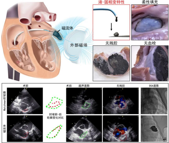
Under ultrasound guidance, the magnetofluid material is injected into the cavity of the left atrial appendage through an interventional catheter. Thanks to its low-viscosity fluid properties, the material can spread naturally along the gaps between the pectinate muscles and into recesses within the cavity, achieving three-dimensional filling of complex chamber structures. Postoperative three-dimensional CT reconstruction and anatomical sections showed no measurable gap between the magnetic gel and the cavity wall, achieving a "zero residual leak" appearance at the imaging level.
Breakthrough 2: A magnetic-field-assisted stabilization mechanism ensures safe shaping
Blood flows rapidly inside the heart, and preventing the liquid material from being dispersed is the core challenge for putting the technique into practice. The team therefore devised an ingenious external "temporary magnetic-field constraint mechanism." In the early stage of injection, an extracorporeal magnetic field provides directional magnetic force, so that the magnetofluid gathers stably within the target area and simultaneously completes in situ solidification. In vitro fluid dynamics experiments showed that, under simulated left atrial blood-flow shear conditions, magnetic-field constraint can provide a safety redundancy threshold roughly six times the scouring force of the blood flow, thereby ensuring that the shaping process is stable and controllable. Once the occluder shape has been fully printed and has solidified and hardened, it generates adhesive and supporting force and can be fixed at the lesion site without an external magnet, at which point the magnetic field is withdrawn.
Breakthrough 3: Dual-mode guidance, safe and universally accessible
The combination of magnetofluids with left atrial appendage occlusion is one of the fruits of ultrasound-guided interventional technology. Radiopaque and ultrasound-imaging components have been added to the magnetofluid material, so it can be visualized under both ultrasound and radiography. The procedure can therefore be performed in the catheterization laboratory of a major hospital, but equally in a grassroots medical institution equipped only with ultrasound, or even on a mobile surgical vehicle, greatly advancing the diffusion of the technology to lower levels of the health system and allowing patients in remote areas to benefit as well.
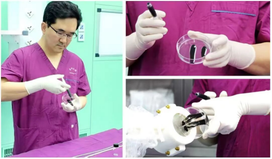
In sum, thanks to its fluidity, solidifiability and magnetic responsiveness, the magnetofluid allows 3D printing technology to play its proper role in cardiac treatment. By combining ultrasound-guided technology with the new magnetofluid material, the team has completely resolved the contradiction of fit between "the soft, beating heart" and "the rigid occluder."
03 Breaking Through Chokepoints and Opening a New Track: Building a Foundation on Methodological Innovation and Generating New Quality Productive Forces Through Disruptive Technology
Academician Hu Shengshou, director of the National Center for Cardiovascular Diseases, recognized the direction in which the discipline was heading and, more than a decade ago, assembled a hybrid technical team that combined the strengths of interventional and surgical approaches, exploring and innovating continuously to develop and progressively refine the ultrasound-guided interventional technology system — laying a solid methodological foundation for this magnetofluid breakthrough.
The appearance of an original Chinese medical device in a top international general journal is not a single academic breakthrough, nor mere persistence, nor simply the accumulation of time. What people see is one device and one paper, but behind it lies the innovation and support of a vast methodology — proving once again that only original, disruptive innovation can create new quality productive forces and give rise to new industries, new models and new drivers of growth:
Ultrasound-guided interventional technology is a methodology; it can continuously generate new technologies and is a form of sustainable innovation. To date, 31 of its technologies have been verified as world firsts, more than 60 domestic and international patents have been obtained, and more than 20 devices have been approved for market in China and the European Union. Among them, the ultrasound-guided guidewire, ultrasound catheter and fully degradable closure device are the only such products on the market worldwide, opening up a new category of medical devices and achieving the major breakthrough of "having what others do not." In 2025 the top medical journal JAMA published the clinical results of a degradable atrial septal defect occluder — the first original Chinese device to appear in JAMA in the journal's 150-year history, breaking the label that Chinese devices are imitative and cheap.
The technology requires neither expensive large imported equipment nor a complex team. It not only solves the "chokepoint" problem but also lowers the technical difficulty, making it possible to train large numbers of grassroots doctors quickly without "cherry-picking the elite," markedly raising grassroots healthcare capability. It has turned cardiac surgery from a "major operation" requiring a trip to a big city and a leading surgeon at a major hospital into an "outpatient procedure," and has even led to the invention of the ultrasound-guided interventional surgical vehicle, which can deliver treatment "from village to village," pioneering a new model of "doorstep surgery" for cardiovascular disease.
More importantly, methodological innovation can bring a "dimensional reduction" attack to bear on traditional technical problems. The new magnetofluid occluder published in Nature is yet another new technology and new product generated by ultrasound-guided interventional technology, demonstrating once again the enormous systemic advantage that methodological innovation brings. Ultrasound-guided interventional technology is like building an entirely new expressway, and there is every reason to believe that more "vehicles" will "change lanes and overtake" on this expressway, making a still greater contribution to human health.

Pan Xiangbin is currently Deputy Director of the National Center for Cardiovascular Diseases and Vice President of Fuwai Hospital, Chinese Academy of Medical Sciences, and a doctoral supervisor in both cardiac medicine and cardiac surgery. He is Director of the Structural Heart Disease Intervention Quality Control Center of the National Health Commission, a standing committee member of the All-China Youth Federation, and a national committee member of the Society of Cardiothoracic and Vascular Surgery of the Chinese Medical Association. He has led more than twenty national and provincial/ministerial projects, including projects under the National Key R&D Program and the National Natural Science Foundation of China.
Source: Fuwai Hospital

